Consultation Hours:

08:30 AM-10:30 AM & 05:00 PM -08:00 PM (Mon-Sun)

Call Us:

+91 93769 93355

Thrombolysis

What is thrombolysis?

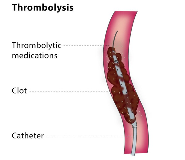
Thrombolysis is a procedure which uses medication to dissolve a blood clot. The medications used in thrombolysis are called thrombolytic agents.
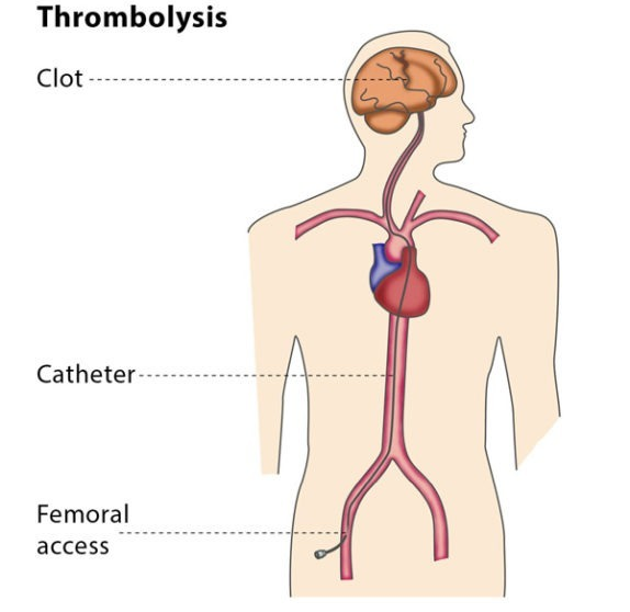
Thrombolysis is most commonly performed to treat a blood clot in the lower limbs, which causes acute limb ischaemia (restricted blood flow in the affected limb), leading to pain in affected area. It can also be used to treat blood clots in organs such as the liver or kidney, as well as treating restricted blood flow in the small intestine due to inflammation or an injury, massive pulmonary embolism (a blood clot in main artery of a lung) or stroke. 
Varicose Veins Doctor in Jaipur, Spider Veins Specialist in Jaipur, Best Thyroid Ablation Specialist in Jaipur, Uterine Fibroid Treatment in Jaipur, Fibroids Surgeon in Jaipur, Dr Manish Rajput, ED Specialist Jaipur

How does the procedure work?

The interventional radiologist will insert a 3 mm plastic tube (called a sheath) into the base of your skull or your groin and will guide the sheath to the blood clot.

Thrombolysis is performed using medications which prevent blood clots from growing (known as lytic substances), which are administered via a catheter into the thrombus. Catheters designed to be used in this treatment have lots of tiny holes. This means that the distribution of lytic substances can be spread out across the catheter and have maximum effect. 
Varicose Veins Doctor in Jaipur, Spider Veins Specialist in Jaipur, Best Thyroid Ablation Specialist in Jaipur, Uterine Fibroid Treatment in Jaipur, Fibroids Surgeon in Jaipur, Dr Manish Rajput, ED Specialist Jaipur

Why perform it?

Thrombolysis is performed to remove the blood clot and to prevent the vein or artery from becoming permanently blocked and restricting blood flow to a limb or organ. Restricted blood flow leads to acute symptoms of pain, a lack of pulse, paleness, paraesthesia (when a limb ‘falls asleep’) and paralysis, as well as the possibility of permanent complications such as tissue necrosis (the death of tissue cells in your body).
You will probably also need medication to prevent blood clots forming as well as a thrombectomy, for which you will be under strict observation for a 24-48 hours with several angiographic follow ups.
You will also be treated for the underlying condition which caused the thrombus, which may be a heart rhythm disorder or a small blood clot caused by deep venous thrombosis.

What are the risks?

Minor risks include the risk of bruising at the puncture site in your neck or groin, or in the affected limb.
The major risks tend to be due to the devices used in the procedure, such as the risk of the blood clot travelling deeper into the artery or vein or an injury to the affected area during the treatment.
In rare cases, the patient experiences bleeding in the skull due to the thrombolysis – if this occurs, the treatment will be stopped immediately.

Bibliography

1. Results of a prospective randomized trial evaluating surgery versus thrombolysis for ischemia of the lower extremity. The STILE trial. Annals of surgery. 1994; 220(3):251-66; discussion 66-8. 2. Berridge DC, Kessel D, Robertson I. Surgery versus thrombolysis for acute limb ischaemia: initial management. Cochrane Database Syst Rev. 2002(3):CD002784. 3. Comerota AJ, Weaver FA, Hosking JD, Froehlich J, Folander H, Sussman B, et al. Results of a prospective, randomized trial of surgery versus thrombolysis for occluded lower extremity bypass grafts. American journal of surgery. 1996; 172(2):105-12. 4. Fox D, Ouriel K, Green RM, Stoughton J, Riggs P, Cimino C. Thrombolysis with prourokinase versus urokinase: an in vitro comparison. Journal of vascular surgery. 1996; 23(4):657-66. 5. Ouriel K, Veith FJ, Sasahara AA. A comparison of recombinant urokinase with vascular surgery as initial treatment for acute arterial occlusion of the legs. Thrombolysis or Peripheral Arterial Surgery (TOPAS) Investigators. The New England journal of medicine. 1998; 338(16):1105-11.

Thrombectomy

What is an endovascular thrombectomy?

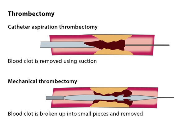
An endovascular thrombectomy is the removal of a thrombus (blood clot) under image guidance. A thrombectomy is most commonly performed for an arterial embolism, which is an arterial blockage often caused by atrial fibrillation, a heart rhythm disorder. An arterial embolism causes acute limb ischaemia (restricted blood supply) which leads to pain in the affected area. A thrombectomy can also be used to treat conditions in your organs, such as in your liver or kidney.
Your doctor may recommend that you have a thrombectomy as a treatment for stroke or for mesenteric ischaemia, where the blood flow in your small intestine is restricted due to inflammation or injury.

How does the procedure work?

The interventional radiologist will insert a 3 mm plastic tube (called a sheath) into the base of your skull or your groin. They will guide the sheath to the blood clot.
There are a number of different techniques for this procedure. The blood clot can be removed using a vacuum to suck the thrombus out, or mechanical equipment to break up the clot, or with clot using saline jets or ultrasound waves.
Varicose Veins Doctor in Jaipur, Spider Veins Specialist in Jaipur, Best Thyroid Ablation Specialist in Jaipur, Uterine Fibroid Treatment in Jaipur, Fibroids Surgeon in Jaipur, Dr Manish Rajput, ED Specialist Jaipur
The devices for these techniques are inserted over the sheath into the affected artery. You may also need additional catheter thrombolysis, a procedure in which clot-dissolving medication is delivered to the clot under X-ray imaging.
A combined treatment using both thrombectomy and thrombolysis can continue over 24-48 hours with several follow-ups using angiography at regular intervals.

Why perform it?

An interventional thrombectomy is used to remove the blood clot and to avoid a permanent blockage in the vein or artery which would prevent blood flow to a limb or an organ, causing acute symptoms of pain, lack of a pulse, paleness, paraesthesia (when a limb ‘falls asleep’) and paralysis, as well as the possibility of permanent complications such as the death of tissue cells in your body.
A thrombectomy is often combined with other treatments, such as treatment to stop blood clots forming or thrombolysis, which involves using medication to break the blood clot down and is followed by strict observation for 24-48 hours.
You will also be treated for the underlying condition which caused the arterial thrombus, such as a heart rhythm disorder or a small blood clot caused by deep venous thrombosis.

What are the risks?

Minor risks include the risk of bruising at the puncture site in the neck or groin or in the affected limb. Major risks include the risk of the blood clot travelling deeper into the artery or vein or an injury to the affected area during the treatment.
In rare cases, patients experience bleeding in the skull as a result of the combined thrombectomy and thrombolysis – if this occurs, the treatment must be stopped immediately.

Bibliography

1. Ansel GM, George BS, Botti CF, McNamara TO, Jenkins JS, Ramee SR, et al. Rheolytic thrombectomy in the management of limb ischemia: 30-day results from a multicenter registry. Journal of endovascular therapy: an official journal of the International Society of Endovascular Specialists. 2002; 9(4):395-402.
2. Allie DE, Hebert CJ, Lirtzman MD, Wyatt CH, Keller VA, Khan MH, et al. Novel simultaneous combination chemical thrombolysis/rheolytic thrombectomy therapy for acute critical limb ischemia: the power-pulse spray technique. Catheterization and cardiovascular interventions: official journal of the Society for Cardiac Angiography & Interventions. 2004; 63(4):512-22.
3. Kasirajan K, Gray B, Beavers FP, Clair DG, Greenberg R, Mascha E, et al. Rheolytic thrombectomy in the management of acute and subacute limb-threatening ischemia. Journal of vascular and interventional radiology: JVIR. 2001; 12(4):413-21.
4. Kasirajan K, Haskal ZJ, Ouriel K. The use of mechanical thrombectomy devices in the management of acute peripheral arterial occlusive disease. Journal of vascular and interventional radiology: JVIR. 2001; 12(4):405-11.
5. Wagner HJ, Muller-Hulsbeck S, Pitton MB, Weiss W, Wess M. Rapid thrombectomy with a hydrodynamic catheter: results from a prospective, multicenter trial. Radiology. 1997; 2 05(3):675-81.